Radius Bone Labelled Diagram / Radius And Ulna Diagram / Radius, in anatomy, the outer of the two bones of the forearm when viewed with the palm facing forward.
byAdmin-
0
Radius Bone Labelled Diagram / Radius And Ulna Diagram / Radius, in anatomy, the outer of the two bones of the forearm when viewed with the palm facing forward.. Study guide for students and teachers. (ii) name the structure labelled a, which attaches muscle to bone. Each bone is a complex living organ that is made up of many cells, protein fibers, and minerals. It is one of the two bones of the forearm, the other being the ulna. The radius is the bone which is present laterally, which means when your palm is facing upwards, it is away from the middle of your body.
The ulna is usually slightly longer than the radius, but the radius is thicker. The skeleton acts as a scaffold by providing support and protection for the soft tissues that make up the rest of the body. In humans it is shorter than the other bone of the forearm, the ulna. Labeled ulna and radius » diagram text anterior labeled ulna and radius anatomy of the ulna and radius bones print options de labeled ulna. In the medial surface, there.
Labelled Diagram Skeleton Teaching Resources from az779572.vo.msecnd.net The radius is the bone which is present laterally, which means when your palm is facing upwards, it is away from the middle of your body. Related posts of human back bones diagram labelled diagram of radius bone. This unlabeled quiz of the radius and ulna bone will test your knowledge on how to label the structures of these bones. Drag and drop the pins to their correct place on the image. The radius and ulna together constitute the forearm. This ulnar view labelled illustration is from 'asklepios atlas of the human anatomy'. The tendon of the brachioradialis. 12 photos of the labelled diagram of radius bone.
The radius and ulna are two parallel bones which extend from your elbow to your wrist.
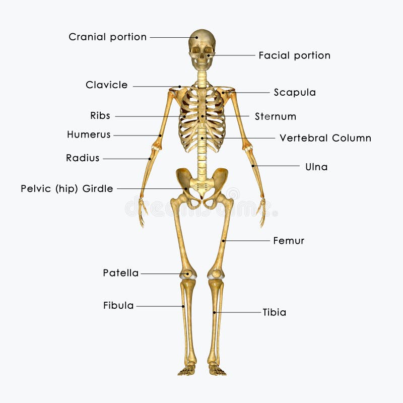
In humans it is shorter than the other bone of the forearm, the ulna. Learn vocabulary, terms and more with flashcards, games and other study tools. Learn the names by coloring. The radius is considered the most commonly fractured bone in the human body, with distal radius fractures being the most common form of radial. This long muscle travels nearly the entire length of the back. You will be required to label the ulnar notch, styloid process of ulna, trochlear notch, proximal radioulnar joint, olecranon process, coronoid process, distal radioulnar joint, etc. Lower jaw (mandible) collar bone. Almost every skeletal muscle works by pulling two or more bones either closer together or further apart. Radius bone is a photograph by asklepios medical atlas which was uploaded on august 3rd, 2016. The tendon of the brachioradialis. The radius is a long bone in the forearm. Illustration with skeleton of human hand ulna, radius and humerus bones. This post discusses social security disability benefits and bone fractures.
Illustration with skeleton of human hand ulna, radius and humerus bones. Drag and drop the pins to their correct place on the image. This post discusses social security disability benefits and bone fractures. Radius bone location (page 1). The radius is considered the most commonly fractured bone in the human body, with distal radius fractures being the most common form of radial.
Skeleton Labelled Stock Illustration Illustration Of Graphic 41527284 from thumbs.dreamstime.com (ii) name the structure labelled a, which attaches muscle to bone. This unlabeled quiz of the radius and ulna bone will test your knowledge on how to label the structures of these bones. This ulnar view labelled illustration is from 'asklepios atlas of the human anatomy'. It extends obliquely downward into a strong, conical projection. Study guide for students and teachers. Radius bone location (page 1). The radius bone is shorter. Learn the names by coloring.
This tutorial covers basic features of the anatomy of the radius and ulna bones.
Its upper concave surface articulates with the. If you or someone you care for has sustained a fracture and are unable to. Individually selectable every part, ideal for learning. Radius along with ulna connects elbow to forearm. The ulna articulates with the trochlea and the radius articulates with the capitulum. Illustration with skeleton of human hand ulna, radius and humerus bones. At the humerus, they articulate with the condyle. Human body bone joint pains anatomy humerus with radius and ulna bones. The radius bone (os radius) supports the lateral (thumb) side of the forearm and the ulna bone (os ulna) supports the medial (little finger) side. The tendon of the brachioradialis. The radius bone is a long horizontal bone present in the forearm and is also called the radial bone. All land vertebrates have this bone. This post discusses social security disability benefits and bone fractures.
Study guide for students and teachers. Radius along with ulna connects elbow to forearm. Diagram of wrist have bone fracture. It lies laterally and parallel to ulna, the second of the forearm bones. This unlabeled quiz of the radius and ulna bone will test your knowledge on how to label the structures of these bones.
Biology Toppers Left Hand Wrist Bone Well Labelled Facebook from lookaside.fbsbx.com 6 3 bone structure anatomy physiology the long bones are those that. (ii) name the structure labelled a, which attaches muscle to bone. 12 photos of the labelled diagram of radius bone. Learn the names by coloring. A graphic shows the bones of the hand, carpals, metacarpals and phalanges. 570 x 737 jpeg 46 кб. I'm not sure of what you mean by bone diagram. It lies laterally and parallel to ulna, the second of the forearm bones.
The lateral side projects distally as the styloid process.
Radius, in anatomy, the outer of the two bones of the forearm when viewed with the palm facing forward. Learn everything about the anatomy of radius and ulna with our articles, video tutorials, labeled diagrams, and quizzes. The radius bone (os radius) supports the lateral (thumb) side of the forearm and the ulna bone (os ulna) supports the medial (little finger) side. Drag and drop the pins to their correct place on the image. The tendon of the brachioradialis. It lies laterally and parallel to ulna, the second of the forearm bones. Bones of the left hand, view from below, labeled in latin. Diagram of wrist have bone fracture. Illustration with skeleton of human hand ulna, radius and humerus bones. A basic human skeleton is studied in schools with a simple diagram. Radius anatomy pictures and information these pictures of this page are about:radius bone location. Radius along with ulna connects elbow to forearm. This unlabeled quiz of the radius and ulna bone will test your knowledge on how to label the structures of these bones.
It extends obliquely downward into a strong, conical projection labelled radius bone. Human body bone joint pains anatomy humerus with radius and ulna bones.